Model Systems
In order to understand the molecular mechanisms leading to brain metastases and to establish new treatment strategies, relevant animal model systems are of importance.
Hovedinnhold
Xenograft models
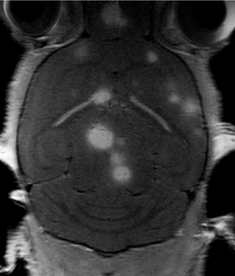
We have developed novel model systems in our lab, where human brain metastasis tumour cell lines from melanoma, lung cancer or human breast cancer are injected into the bloodstream of nod/scid mice. Through a highly standardized procedure, we inject the cell lines into the left cardiac ventricle in mice, and study tumour development by using multimodal, molecular imaging. During 3-6 weeks, all animals develop new tumours with a profound preference to the brain. MRI shows multiple metastases within the brain, similar to what is seen in patients (see figure). The models are thus among the very few which, in a systematic and consistent manner, can mimic the metastatic spread of cancers seen in patients.
Patient biobanks
In our studies, we make use of an ethically approved patient biobank, where brain metastasis and blood samples from patients are continuously collected and stored. The biobank is a valuable contribution to our research, and we will use the patient samples to study whether we can find genetic mutations that may be driving metastasis to the brain. By this, we hope to increase our molecular understanding, which can be used for future design of new treatment strategies.
For more information on animal model systems, you can read this publication.
