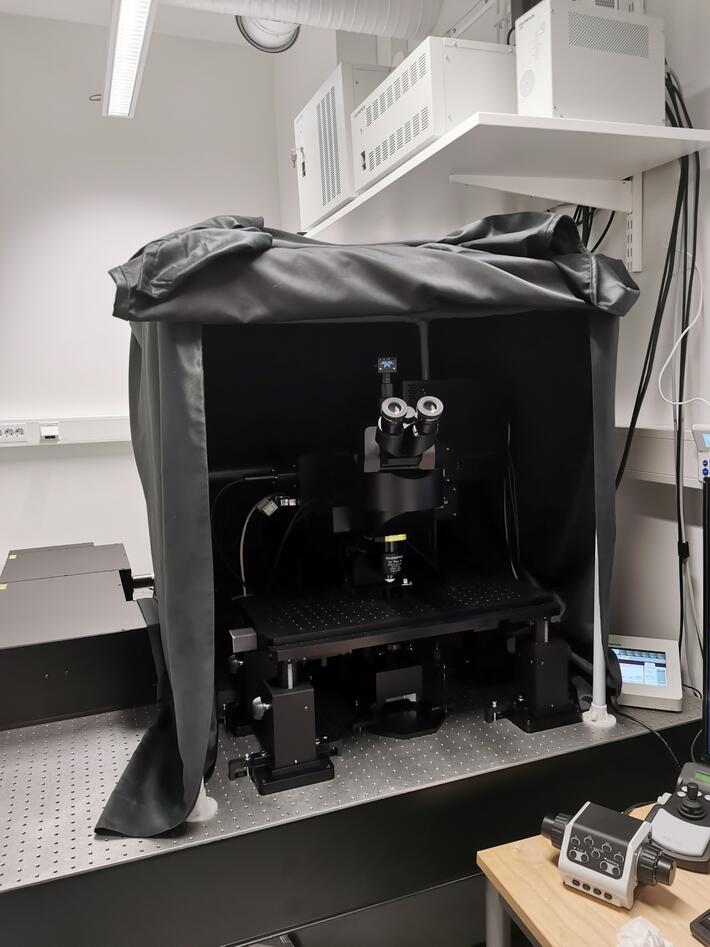
Ex vivo and in vivo multiphoton imaging
Olympus Multiphoton FVMP-RS
Hovedinnhold
Room: 3029, 3rd floor Vivarium (HUS).
Responsible contact persons: Hege Dale, Emmet MC Cormack
FluoView FVMPE-RS laser scanning multiphoton:
- FVMPE-RS hybrid scan unit with standard point scanning and resonant galvanometer scanners.
- 2 non-descanned GaAsp-detectors (NDD) => simultanous two-color imaging. Possible color combinations: Blue (455-490nm)/Green-Yellow (515-560) emission and Green (495-540nm)/Red (575-645) emission.
- Pulsed laser: Mai Tai DeepSee HP (Spectra Physics), an Ultrafast Ti:Sapphire Laser with Automated Dispersion Compensation. Tuning range: 690-1040 nm. The laser is specifically tailored to the FVMPR-RS sytem.
- Objective: 25x NA1.05 Olympus (XPLN25XWMP2) with motorized correction collar and 2 mm working distance. Max field of view 505x505 um. Dip-in water immersion.
- Gantry frame: Large and flexible stage area suited for both live animals, ex vivo and fixed samples.
https://www.sciencemag.org/features/2019/03/shedding-light-deep-tissue-multiphoton-microscopy
Typical samples for multiphoton imaging:
- Ex vivo models; explant tissue, organoids, spheroids.
- 3D-cell culture models.
- Intravital imaging on living animals through an implanted imaging window or chamber.
- Thick fixed samles.
Dependent on the texture of the tissue, the real penetraion depth can vary a lot. Many tissue types are highly light scattering, and this has a negative impact on image acquisition in the depth. Tissue clearing can be a solution, however not necessarily straight forward.
For a nice overview on tissue clearing: Tainaka et al. Cell Reports 2018. Chemical Landscape for Tissue Clearing Based on Hydrophilic Reagents
14.04.2023
