"Google map" for tumors
The University of Bergen has just implemented the Hyperion Imaging System, next generation immunohistochemistry where researchers can explore tissue biology with 35 antibodies simultaneously. This instrument is the only of its kind in Northern Europe, and is open for booking for all researchers on equal terms.

Main content
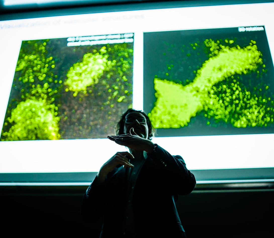
The Hyperion was recently introduced in a grand opening symposium, which attracted great interest in our research communities. Leigh-Anne McDuffus from the Fluidigm Corporation explained the features and possibilities of the Hyperion Imaging System. Keynote speaker was Dr. Dario Bressan, Head of the IMAXT Laboratory, CRUK Cambridge Institute, University of Cambridge, UK.
The Hyperion can help us to fully map tumors, providing extraordinary precision.
"The Hyperion Imaging System can, in a way, be compared to a very powerful microscope, which makes it possible to study cancer cells and create a molecular map of the tissue. In this way, we can see more clearly how cancer cells and tissue around the cells work, and the hope is that in the future, we can tell which patients have aggressive tumors and which have less dangerous tumors", explains CCBIO Director Professor Lars A. Akslen in a video posted on Youtube.
CCBIO is the formal owner of the new analyzer that was presented Wednesday 6 February. Many partners are behind this, and in addition to CCBIO, the Faculty of Medicine and the Bergen Research Foundation (BFS) have contributed financially. Access to the equipment is managed and operated by the Flow Cytometry Core Facility.
The first of its kind in Northern Europe
This multiplexing tissue analyzer is the only one of its kind in Norway and in Scandinavia, and to date also in Northern Europe, and is available for use by both national and international researchers. Dean of Research at the Faculty of Medicine, Marit Bakke, hopes that the instrument can stimulate even more collaboration:
"I hope this new core facility will provide a breeding ground for new collaborative projects between the hospital and the Faculty of Medicine", Bakke declared, at the symposium which was arranged in honor of the new acquisition.
Facilitates excellent research
CCBIO’s Administrative Leader Geir Olav Løken is happy to invite all interested researchers to use the instrument. "The Hyperion will be organized as a core facility. All researchers can use the instrument on a pay-per-service basis on equal terms without demand for co-authorship".
Head of Department at the Department of Clinical Medicine, Kjell-Morten Myhr, believes that the equipment is a very positive factor for the research environment in and around the University of Bergen:
"The analyzer offers opportunities to produce top research in Bergen, and opportunities for research collaboration, both nationally and internationally," said Myhr, and, like others, emphasized the large common effort behind the acquisition, and the organization of the core facility.
Sveinung Hole, CEO of Bergen Research Foundation, was happy to support the establishment of this unique and cutting edge technology at the medical campus in Bergen.
Creating new research possibilities
The Hyperion Imaging platform is combining two modules: the Hyperion Tissue Imager and a Helios CyTOF® system. The Helios/Hyperion system use metal tagged antibodies towards specific proteins of interest in select regions of fixed tissue sections, frozen tissue section or cell smears or cell suspension.
This technology utilize a state-of-the-art Time-of-Flight Inductively Coupled Plasma mass spectrometry technology together with elemental tags that have higher molecular weights than those elements that are naturally abundant in biological systems.
The advantage with using mass cytometry on cells in suspension is that it does not produce the same overlap as when using fluorochromes in flow cytometry, and thus there is little need for compensation. No fluorescence background also makes this technology very suitable for looking at high dimensional functional and phenotypic correlations at single cell level, accelerating biomarker findings and developing targeted drug therapy (precision medicine).
The Hyperion image system provides a visual context to the heterogenisity of tissue microenvironemnts we have not been able to do before. This is possible because of the up to 37 metal tags used to simulataneously detect multiple proteins on one tissue section, which alowes for understanding of protein behaviors and interactions to drive biological breakthroughs and define clinical biomarkers.
Or very short said, as key speaker Dr. Dario Bressan from the University of Cambridge used as an analogue, consider it a "google map" for tumors!
For more information on research possibilities, use this link.
