Decoding Sensory Navigation: Insights into Larval Settlement Mechanisms in Marine Invertebrates
A new paper from the Chatzigeorgiou Group unravels the enigmatic sensory strategies of planktonic larvae using Ciona intestinalis as a model organism.

Main content
Among the most abundant organisms inhabiting the oceans are numerous marine invertebrates with biphasic life cycles. Their freely-swimming larvae disperse in the water column in search of a site where they can attach and undergo a transformative process termed metamorphosis, giving rise to benthic sessile adults. One of the biggest challenges for these diminutive creatures lies in their ability to detect the relevant chemical and mechanical cues that will guide them to suitable settlement sites. How they achieve this remains a mystery.
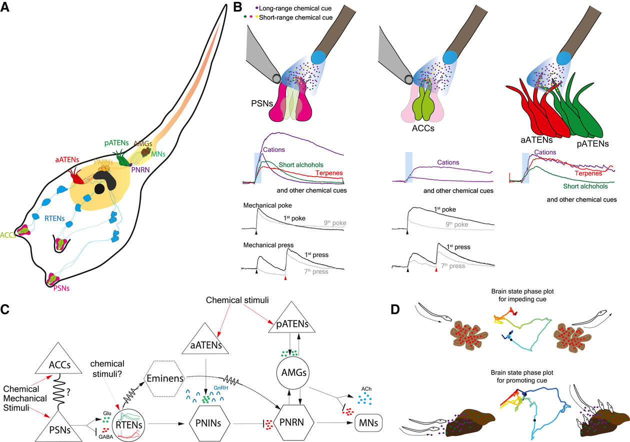
In a recent study published in Current Biology, researchers in the Chatzigeorgiou lab led by Chief Engineer Jørgen Høyer used the genetically tractable model organism Ciona intestinalis to solve this enigma. Through functional imaging assays, they discovered that a small number of sensory cells in the animal's head could detect various mechanical stimuli as well as a surprising diversity of long-range and short-range chemical cues abundant near the sea floor. This discovery revealed that Ciona leverages polymodal sensory cells to map its surrounding sensory environment.
“We used a technique called calcium imaging to study the neuronal activity in our model organism Ciona, an animal well suited for this approach," explained Jørgen. "With this technique, we can watch and follow the neurons fire in the moment. From the activation of the sensory cells that monitor the environment, to the brain and further downstream to the neurons that control motion.”
They showed that different chemical and mechanical cues elicit qualitatively and quantitatively distinct response profiles in the same sensory cells, suggesting that sensory data is already encoded at the periphery of the nervous system. This ability allows planktonic larvae with streamlined nervous systems to sense and encode diverse stimuli effectively.
The researchers hypothesized that both short and long-range chemical cues held significant ecological and ethological importance. To examine this hypothesis, they designed quantitative behavioral assays to assess the impact of these cues on settlement and metamorphosis behaviors. Their findings revealed that several chemical cues strongly influenced in either a positive or negative manner the rates at which larvae attached to the substrate and underwent metamorphosis.
The hope is that our results contribute to the understanding of the sensory system of not only Ciona larvae but also other marine life as there are many animals in the ocean with a biphasic life cycle, starting out as larvae.
-Jørgen Høyer
The research team's collaborative efforts integrated a diverse range of skills, encompassing both software development and experimental assays. The project was initiated by former Department Engineer Kushal Kolar, currently pursuing a PhD at NYU, who was curious about the sensory ecology of planktonic larvae. During his tenure in the Chatzigeorgiou Lab, Kushal established mechanical stimulation and chemical perfusion calcium imaging assays. “His calcium imaging analysis software (Mesmerize) was critical for us to be able to carry out complex calcium imaging analysis,” noted group leader Marios Chatzigeorgiou. Athira Athira developed the computational tools to interpret the intricate signaling dynamics observed during whole-brain imaging experiments, while intern Meike van den Burgh conducted numerous behavioral assays, contributing to the understanding of how chemical cues influence larval attachment and metamorphosis.
By employing cutting-edge functional imaging techniques, the researchers captured high-speed neuronal dynamics across the entire brain. They observed that even at rest, Ciona larvae displayed neuronal activity in localized brain regions that were likely required for spontaneous locomotion. Moreover, the presentation of attractive or repulsive cues led to drastically different brain-wide activity patterns, suggesting that the streamlined nervous system of Ciona can compute the valence (attractive or repulsive) of a particular chemical cue. This process then likely activates specific neuronal pathways, guiding the larvae to make stimulus-driven behavioral action choices, such as whether to stay or leave.
Reflecting on the project, Marios highlighted Jørgen’s contribution to the findings. “He is an electrical engineer by training and I was truly impressed by his ability to get out of his comfort zone to perform transgenesis experiments and to carry out highly demanding calcium imaging assays,” said Marios. Jørgen added, “I felt it was a big opportunity to learn something new. It’s not every day that I get the chance to take the lead on experiments in a biology project. And it's been very interesting to look inside the head of these animals and actually see what's going on. The hope is that our results contribute to the understanding of the sensory system of not only Ciona larvae but also other marine life as there are many animals in the ocean with a biphasic life cycle, starting out as larvae."