Improved understanding of oxygen sensors in human cells
Oxygen is vital for human life and so is our ability to respond to low oxygen levels. Researchers at the University of Oxford and University of Bergen have uncovered new factors defining how human cells respond to hypoxia.

Hovedinnhold
One of the most well-studied mechanisms by which animals achieve oxygen homeostasis is via the hypoxia-inducible factor (HIF) transcription factors. However, five years ago, another key player in oxygen sensing was discovered by researchers at the University of Oxford: the human ADO enzyme (2-Aminoethanethiol dioxygenase, or cysteamine dioxygenase) (Science, 2019). ADO regulates the stability of proteins in an O2-dependent manner via oxidation of Cysteine-starting proteins which leads to their cellular degradation. ADO-mediated control of protein stability therefore has the potential to be a significant regulator of physiological responses to hypoxia, in particular to provide a rapid response to hypoxia, compared to regulation via the HIF system which initiates hypoxic adaption by increasing gene expression.
Three protein substrates of ADO were previously characterized: regulators of G-protein signalling 4 and 5 (RGS4/5), and chemokine interleukin-32 (IL32), whose stability were ADO- and O2-dependent. However, there are 200-300 human proteins starting with a Cysteine (i.e. having Cysteine at the protein N-terminus after co-translational Methionine cleavage). All of these represent potential human ADO substrates that could be hypoxically regulated. The question therefore arises as to how many and which proteins are substrates of ADO and thus potentially involved in hypoxic responses.
Teams in the laboratories of Emily Flashman and Peter Ratcliffe (University of Oxford) screened a variety of Cys-starting proteins in vitro and in human cells. ADO was found to be selective for those proteins with certain amino acid residues following the N-terminal Cysteine (basic or aromatic residues). Interestingly, some proteins were unexpectedly not regulated by ADO in cell-reporter assays despite being good substrates in vitro, raising the possibility that their N-termini were protected from oxidation in human cells. A plausible explanation could be that they were modified by other chemical groups. The most common protein modification occurring on the N-terminal end of human proteins is N-terminal acetylation.
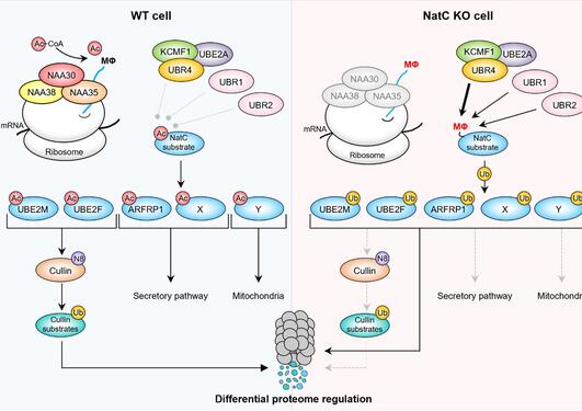
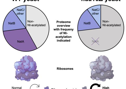
Based on many years of research defining this modification and the responsible enzymes, the group of Thomas Arnesen at the Department of Biomedicine, University of Bergen, was invited to join these efforts. UiB researcher Matti Myllykoski, PhD-student Malin Lundekvam and postdoctor Nina McTiernan performed a number of experiments using purified enzymes, synthetic peptides, human cells as well as the classical model organism budding yeast (Saccharomyces cerevisiae). The conclusion was clear: Proteins with Cys-starting N-termini could be N-terminally acetylated by an enzyme called NatA. Furthermore, the preference (substrate specificity) of NatA was totally different to what was observed for ADO. NatA was selective among Nt-Cys sequences in vitro, with a preference for acidic or polar residues immediately following the Nt-Cys. Despite these opposite preferences of NatA and ADO, some “intermediate” proteins were potential substrates of both ADO and NatA. However, further experiments revealed that N-terminal acetylation and N-terminal oxidation are mutually exclusive modifications: one will block for the other.
These data suggest that NatA and ADO have evolved to mostly modify distinct pools of cellular Cys-starting proteins. The ADO substrate proteins are degraded during normal oxygen levels while the NatA substrates are stable. The proteins potentially targeted by both NatA and ADO may be degraded via ADO during conditions which may reduce NatA activity such as stress or disease.
In sum, this study helps define which proteins are available for ADO and O2-dependent proteostatic regulation, and thereby contributes to our understanding of oxygen sensing in human cells.
The published article is found here.


