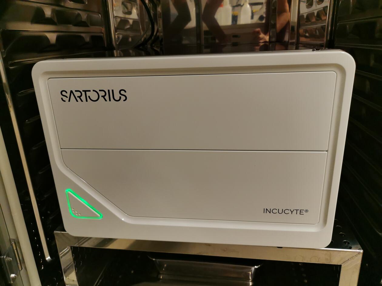
IncuCyte S3 - Live cell analysis
Main content
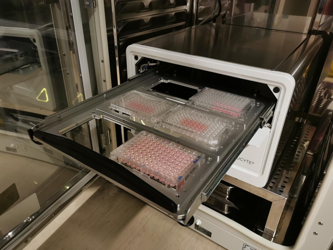
The Sartorius IncuCyte S3 Live-Cell Analysis System arrived at MIC early 2022 and is a high throughput system for imaging primary over several days (2-5 days typically but longer also possible). It is placed inside a cell incubator for optimal cell-growth conditions. Examples of studies on cell cultures; proliferation, viability, cell migration and invasion, apoptosis and cytotoxicity. Imaging and analysis of spheroids and organoids are also possible.
- Objectives: 4x, 10x and 20x.
- Green and red fluorescence, brightfield and phase contrast.
- Low resolution -> high throughput.
- Up to 6x 96 well plates simultaneously, but flexible vessel geometry.
- Wound-maker for 96 well plates for wound healing assays.
- Running protocols suitable for many types of experiments.
- Possibilities for multi-users and multi-experiments running simultaneously.
- Possible to simultanounsy run protocols with different time scheduling and different objectives between plate positions.
Training by MIC personnel is mandatory before starting experiments. Booking of the instrument is compulsory and you have to thoroughly familiarize yourself with the instrument guidelines and rules, particularily to avoid spoiling already running experiments.
- Please download the pdf IncuCyte_S3_user manual_MIC under documents at the bottom of this page.
The method for analysing the data must be optimized for each type of experiment, and analysis of the data can be performed progressively or after finalizing the acquisition (recommended).
Software modules for analysis available on our system:
- Cell Migration
- Single and multi Spheroid imaging
- Organoid (embedded in martigel) imaging
Under documents (below) there are several well described protocols on relevant applications (made by Sartorius).
More info: https://www.sartorius.com/en/products/live-cell-imaging-analysis/live-cell-analysis-instruments

