Microdissection of tissue and cells
Laser Microdissection System
Main content
Leica LMD7
Location: Department of Pathology, 2nd floor, Sentralblokken, Haukeland University Hospital, room 5322 (AV-room)
Contact person: Kenneth Finne
Laser microdissection enables the user to isolate areas of tissue, or even single cells, from FFPE (formalin-fixed, paraffin-embedded) or frozen tissue sections. The LMD7 comes with a high-quality Leica microscope and camera, and allows for either brightfield or fluorescent imaging. Laser-microdissected tissue is very suitable for downstream genomic, transcriptomic, proteomic or metabolomic analyses.
Key features:
- High-quality Leica microscope
- High powered laser (cuts through almost “everything”)
- Gravity-assisted tissue isolation
- Four air-objectives with 5x, 10x, 40x and 63x magnification
- Fluorescence filters (RGB and Cy5)
- Several different collection alternatives (tube caps, 96-well plates, 8/12-tube strips)
- User-friendly software
Read more here: https://www.leica-microsystems.com/products/light-microscopes/p/leica-lmd7/
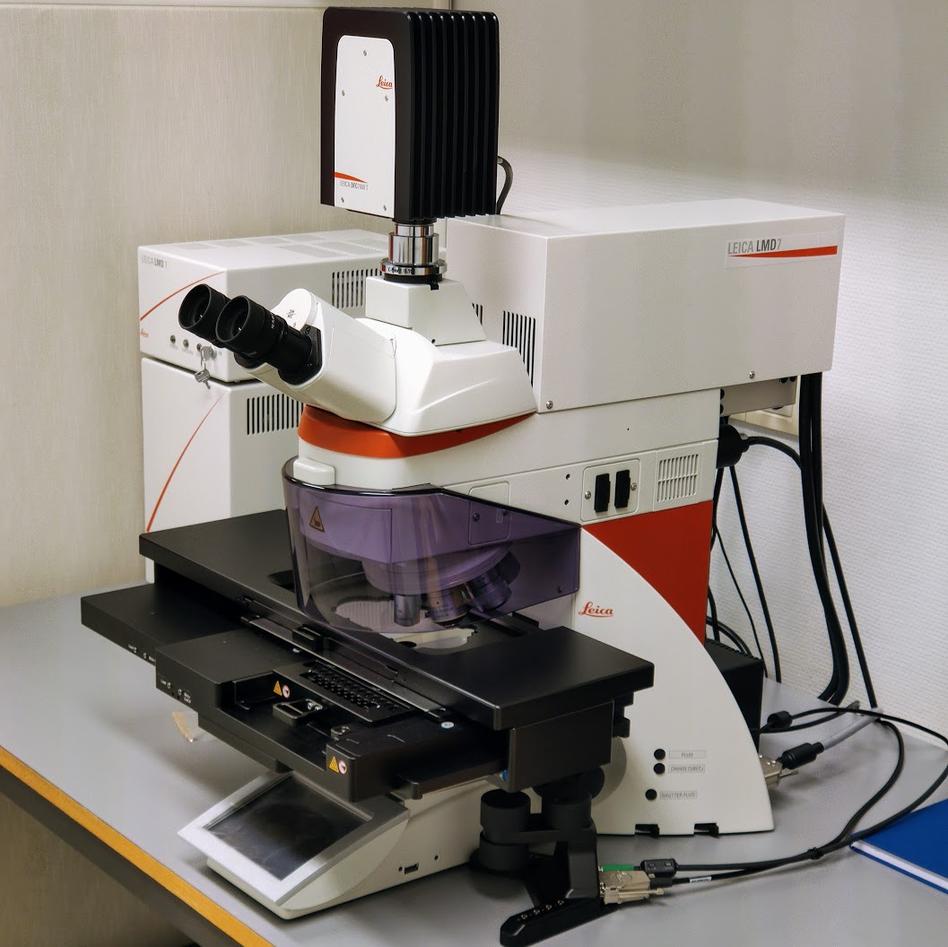
Photo:
Kenneth Finne
03.09.2020